Research Abstract

ヒトiPSC由来の最適化された心筋細胞を用いることで、心臓での生着、増殖、治療可能性を向上させる

Enhanced engraftment, proliferation, and therapeutic potential in heart using optimized human iPSC-derived cardiomyocytes

2016年1月8日 Scientific Reports 6 : 19111 doi: 10.1038/srep19111

ヒトiPSC由来の最適化された心筋細胞を用いることで、心臓での生着、増殖、治療可能性を向上させる
拡大する

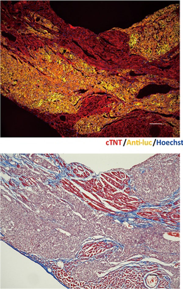
ヒト多能性幹細胞由来の心筋細胞(CM)は心臓の細胞治療のための有望なツールである。誘導多能性幹細胞(iPSC)由来のCMの移植がいくつかの動物モデルで報告されているが、その治療効果は限定的であり、おそらく移植された細胞が十分に最適化されていなかったためではないかと考えられる。心臓の再構築に用いる移植細胞を最適化するために、未分化iPSC、分化誘導開始後4日目の中胚葉細胞、8日目、20日目、30日目の純化したiPSC-CMを心筋内に移植し、in vivo光イメージングを用いて生着率(ER)の変化を追跡することで、それらの細胞の生着効率の比較を行った。この解析から、20日目CMのERが他の細胞より有意に高いことが明らかになった。免疫不全マウスの梗塞心臓へ20日目CMを移植すると生着が良好であることが示され、また心エコー検査から細胞治療による有意な機能改善が示された。そのうえ、移植後3カ月の時点までのin vivo光イメージングのシグナルやKi67陽性CMの比率から、生着したCMは宿主の心臓で増殖していることが示された。この移植組織の増大は3カ月でプラトーに達するが、組織学的解析から移植細胞の成熟は3~6カ月まで進むことが確認された。これらの結果から、20日目CMは宿主マウス心臓での生着、増殖、治療能が非常に高いと考えられた。また、このモデルは長期間にわたり移植細胞の運命を追跡するのに使用できることが実証された。

Shunsuke Funakoshi, Kenji Miki, Tadashi Takaki, Chikako Okubo, Takeshi Hatani, Kazuhisa Chonabayashi, Misato Nishikawa, Ikue Takei, Akiko Oishi, Megumi Narita, Masahiko Hoshijima, Takeshi Kimura, Shinya Yamanaka & Yoshinori Yoshida

Corresponding Author

吉田 善紀
京都大学iPS細胞研究所

Human pluripotent stem cell-derived cardiomyocytes (CMs) are a promising tool for cardiac cell therapy. Although transplantation of induced pluripotent stem cell (iPSC)-derived CMs have been reported in several animal models, the treatment effect was limited, probably due to poor optimization of the injected cells. To optimize graft cells for cardiac reconstruction, we compared the engraftment efficiency of intramyocardially-injected undifferentiated-iPSCs, day4 mesodermal cells, and day8, day20, and day30 purified iPSC-CMs after initial differentiation by tracing the engraftment ratio (ER) using in vivo bioluminescence imaging. This analysis revealed the ER of day20 CMs was significantly higher compared to other cells. Transplantation of day20 CMs into the infarcted hearts of immunodeficient mice showed good engraftment, and echocardiography showed significant functional improvement by cell therapy. Moreover, the imaging signal and ratio of Ki67-positive CMs at 3 months post injection indicated engrafted CMs proliferated in the host heart. Although this graft growth reached a plateau at 3 months, histological analysis confirmed progressive maturation from 3 to 6 months. These results suggested that day20 CMs had very high engraftment, proliferation, and therapeutic potential in host mouse hearts. They also demonstrate this model can be used to track the fate of transplanted cells over a long time.

「おすすめのコンテンツ」記事一覧へ戻る

プライバシーマーク制度