Research Abstract
波長1030 nmのピコ秒パルスレーザーを用いて、生体用2光子顕微鏡法による海馬ニューロンを可視する
Visualizing hippocampal neurons with in vivo two-photon microscopy using a 1030 nm picosecond pulse laser
2013年1月24日 Scientific Reports 3 : 1014 doi: 10.1038/srep01014
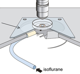
生体用2光子顕微鏡法は脳表面から数百マイクロメートル以上の深部での事象を可視化することは困難であるものの、脳機能にとって不可欠な神経活動の情報を明らかにしてきた。今回、半導体レーザーをベースとする波長1030 nmの新規光源を開発した。これは、出力強度2 W、繰り返し周波数20 MHzで、パルス幅5ピコ秒の光パルスを発生可能である。また、正立顕微鏡ステージにマウス頭部を固定する水平制御機構付きのシステムもあわせて開発した。H-lineマウスの脳を撮像し、深部到達度を検討した結果、この新しいレーザーを用いることで、非常に高い信号背景比で皮質全層に広がる錐体ニューロンを撮像することに成功したのみならず、若齢マウスでは海馬CA1ニューロンをも撮像することが可能あることが示された。
川上 良介1, 2, 5, 澤田 和明1, 2, 佐藤 綾耶3, 5, 日比 輝正1, 2, 5, 小澤 祐市4, 5, 佐藤 俊一4, 5, 横山 弘之3, 5 & 根本 知己1, 2, 5
- 北海道大学 電子科学研究所
- 北海道大学大学院 情報科学研究科
- 東北大学 未来科学技術共同研究センター
- 東北大学 多元物質科学研究所
- 科学技術振興機構 戦略的創造研究推進事業(JST-CREST)
In vivo two-photon microscopy has revealed vital information on neural activity for brain function, even in light of its limitation in imaging events at depths greater than several hundred micrometers from the brain surface. We developed a novel semiconductor-laser-based light source with a wavelength of 1030 nm that can generate pulses of 5-picosecond duration with 2-W output power, and a 20-MHz repetition rate. We also developed a system to secure the head of the mouse under an upright microscope stage that has a horizontal adjustment mechanism. We examined the penetration depth while imaging the H-Line mouse brain and demonstrated that our newly developed laser successfully images not only cortex pyramidal neurons spreading to all cortex layers at a superior signal-to-background ratio, but also images hippocampal CA1 neurons in a young adult mouse.