定量的活動依存性マンガン造影MRIで判定されるマウスのパーキンソン病重篤度
Quantitative activation-induced manganese-enhanced MRI reveals severity of Parkinson’s disease in mice
2015年8月10日 Scientific Reports 5 : 12800 doi: 10.1038/srep12800
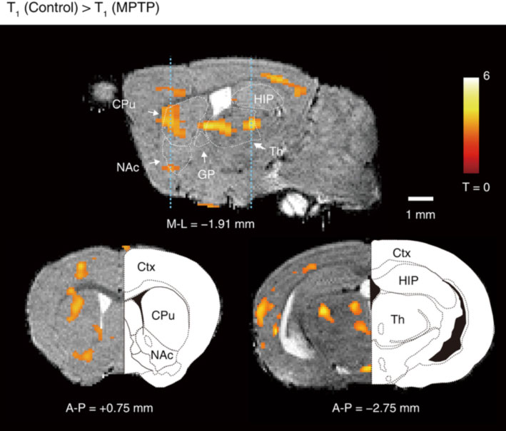
今回マウスで、縦緩和時間 (T1) の定量計測を組み合わせた活動依存性マンガン造影磁気共鳴イメージング法(activation-induced manganese-enhanced MRI with quantitative T1 measurement; qAIM-MRI)により、パーキンソン病(PD)の重篤度を判定できることを示した。まず、ニューロンの活動に依存してマンガンイオンの蓄積が起こることを示した。次に、PDモデルマウスにqAIM-MRI を適用したところ、PDにより神経活動が強くなった大脳基底核の領域は主に尾状核/被殻の背側部であり、その神経活動の強さはPDの重篤度と有意に相関していた。大脳基底核以外では、感覚運動皮質及び、視床の parafascicular nucleus の神経活動が PD の重篤度と高い相関を示した。これらの結果は、PDの症状発現にこれらの領域が関与していることを示唆している。この結果は、PD の発症機序解明に大きな前進をもたらす。また、qAIM-MRI は、血流の変化に左右されず、自由行動下の神経活動を直接計測できる非侵襲計測法であるため、PD の診断法として発展する可能性があるだけでなく、他の脳・神経疾患の病態生理学研究などに応用することができる。
Satomi Kikuta, Yukiyo Nakamura, Yukio Yamamura, Atsushi Tamura, Noriyasu Homma, Yuchio Yanagawa, Hajime Tamura, Jiro Kasahara & Makoto Osanai