Research Abstract
動物脳での回路イメージングによって識別される後部下側頭皮質内のフィードフォワードニューロンと内部投射ニューロン
Distinct Feedforward and Intrinsic Neurons in Posterior Inferotemporal Cortex Revealed by in Vivo Connection Imaging
2012年12月6日 Scientific Reports 2 : 934 doi: 10.1038/srep00934
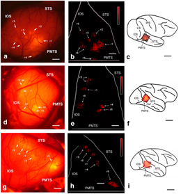
視覚対象の認識にかかわる回路について、マカクザルの下側頭葉前部(TE)および後部(TEO)を、2段階の生体内脳標識法を利用して検討した。まず、TEO内のフィードフォワードニューロンの斑状分布を可視化して次段階の標的とするため、TEに赤色蛍光トレーサーを注入した。次に、その斑状分布を生体脳の皮質表面で可視化し、その斑の1つに緑色蛍光トレーサーを注入した。組織学的検索によれば、TEO内の注入部位から500 μm以上離れた斑には、緑色のTEO内在性の投射ニューロンと赤色のTEOからTEに投射するニューロンが混じり合っていたが、両色素で標識されるニューロンはほとんど見られなかった。それに対し、TEOの注入部位に近い斑には、両色素で標識されるニューロンが多数含まれていた。したがって、TEOには、次の2つの回路が、平行かつ空間的に混じり合った形で分布していると考えられる:(1)非常に限局した側枝を持ち、TEに投射するTEOニューロン、(2)TEO内回路網に広く投射するが、TEには投射しないTEOニューロン。これらの平行システムは、それぞれ速い信号伝達と高度処理後の信号伝達を担っている可能性がある。
一戸 紀孝1, 2, Elena Borra2 & Kathleen Rockland2
- 独立行政法人 国立精神・神経医療研究センター 神経研究所 微細構造研究部
- 独立行政法人 理化学研究所 脳科学総合研究センター 脳皮質機能構造研究チーム
We investigated circuits for object recognition in macaque anterior (TE) and posterior inferotemporal cortex (TEO), using a two-step method with in vivo anatomical imaging. In step 1, red fluorescent tracer was injected into TE to reveal and Pre-target patches of feedforward neurons in TEO. In step 2, these were visualized on the cortical surface in vivo, and injected with green fluorescent tracer. Histological processing revealed that patches >500 μm from the injection site in TEO consisted of intermingled green TEO intrinsically projecting neurons and red TEO-to-TE neurons, with only few double-labeled neurons. In contrast, patches near the injection site in TEO contained many double-labeled neurons. Two parallel, spatially intermingled circuits are suggested: (1) TEO neurons having very local intrinsic collaterals and projection to TE (2) TEO neurons projecting more widely in the intrinsic network, but not to TE. These parallel systems might be specialized for, respectively, fast vs. highly processed signals.