原子間力顕微鏡法による水のネットワークの超高分解能イメージング
Ultrahigh-resolution imaging of water networks by atomic force microscopy
2017年2月3日 Nature Communications 8 : 14313 doi: 10.1038/ncomms14313
金属表面上で成長した水分子の層における局所的な欠陥は、表面が濡れる過程において大きな影響を及ぼす。しかし、そのようなマイナーな構造は巨視的な測定方法では検出することができない。今回我々は、4.8 Kにおける分子修飾探針を用いた非接触原子間力顕微鏡(AFM)によって、銅(110)表面上の水単分子層を極めて高い分解能でイメージングできることを実証する。一酸化炭素を先端に付けた探針によるAFM測定では、水素原子の寄与は小さく、主に酸素原子を反映した像が得られた。この酸素骨格のAFM像によって、この水のネットワークは、局所的な欠陥や末端も含めて、5員環と6員環から構成されていることが明らかになった。今回の結果により、弱く結合した分子集合体の原子構造を決定するための手法としてAFMが有用であることが強く示された。
Corresponding Author
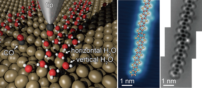
Local defects in water layers growing on metal surfaces have a key influence on the wetting process at the surfaces; however, such minor structures are undetectable by macroscopic methods. Here, we demonstrate ultrahigh-resolution imaging of single water layers on a copper(110) surface by using non-contact atomic force microscopy (AFM) with molecular functionalized tips at 4.8 K. AFM with a probe tip terminated by carbon monoxide predominantly images oxygen atoms, whereas the contribution of hydrogen atoms is modest. Oxygen skeletons in the AFM images reveal that the water networks containing local defects and edges are composed of pentagonal and hexagonal rings. The results reinforce the applicability of AFM to characterize atomic structures of weakly bonded molecular assemblies.