Research Abstract
分泌準備状態にあるSNARE複合体のシナプス前終末と膵β細胞での相違を2光子蛍光寿命画像法を用いて可視化
Two-photon fluorescence lifetime imaging of primed SNARE complexes in presynaptic terminals and β cells
2015年10月6日 Nature Communications 6 : 8531 doi: 10.1038/ncomms9531
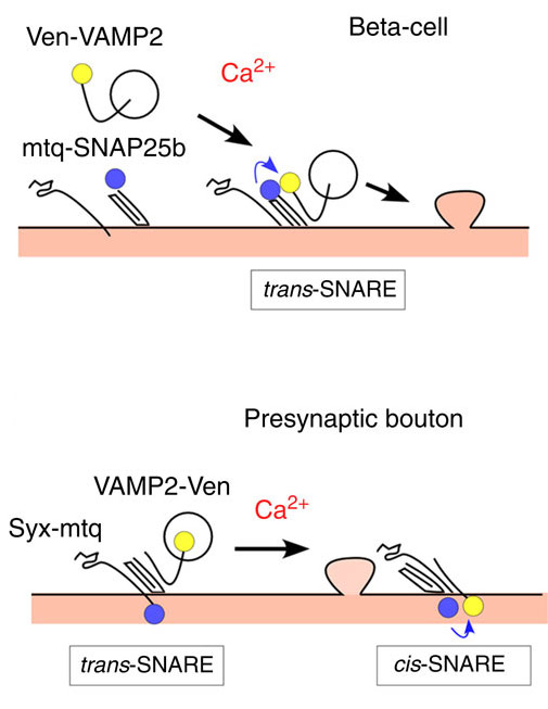
Ca2+依存性の開口放出の速度がSNARE複合体の複合化状態にどのように依存しているのかは明らかになっていない。今回我々は、シナプス前終末中とランゲルハンス島内のβ細胞においてSNARE分子間の複合化で起こる蛍光共鳴エネルギー移動(FRET)を、2光子蛍光寿命画像法(FLIM)を用いて直接測定した。変異動物を用いた実験から、我々の構築したSNAREのプローブは、神経で超高速の開口放出を起こすことが確認された。シナプス前終末では開口放出の準備状態であるtrans-SNARE複合体は活性領域に集積しており、個々の小胞に結合している複合体の数も推定された。対照的に、開口放出がゆっくりと進行するβ細胞では、SNAREはほとんど複合化しておらず、複合化はインスリン開口放出の少し前に起きた。従って、融合速度のちがいはSNARE複合化の度合いで説明されることが示された。SNAREのFRET/FLIM法は固定された組織でも複合化を検出でき、小胞融合の準備状態を生きた組織や固定した標本で可視化することができる。
Noriko Takahashi, Wakako Sawada, Jun Noguchi, Satoshi Watanabe, Hasan Ucar, Akiko Hayashi-Takagi, Sho Yagishita, Mitsuyo Ohno, Hiroshi Tokumaru & Haruo Kasai
Corresponding Author
It remains unclear how readiness for Ca2+-dependent exocytosis depends on varying degrees of SNARE complex assembly. Here we directly investigate the SNARE assembly using two-photon fluorescence lifetime imaging (FLIM) of Förster resonance energy transfer (FRET) between three pairs of neuronal SNAREs in presynaptic boutons and pancreatic β cells in the islets of Langerhans. These FRET probes functionally rescue their endogenous counterparts, supporting ultrafast exocytosis. We show that trans-SNARE complexes accumulated in the active zone, and estimate the number of complexes associated with each docked vesicle. In contrast, SNAREs were unassembled in resting state, and assembled only shortly prior to insulin exocytosis, which proceeds slowly. We thus demonstrate that distinct states of fusion readiness are associated with SNARE complex formation. Our FRET/FLIM approaches enable optical imaging of fusion readiness in both live and chemically fixed tissues.