Research Abstract
CEPIAを使って細胞小器官内のCa2+を細胞レベル以下の分解能で画像化する
Imaging intraorganellar Ca2+ at subcellular resolution using CEPIA
2014年6月13日 Nature Communications 4 : 4153 doi: 10.1038/ncomms5153
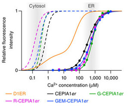
小胞体とミトコンドリアはその内腔にCa2+を取り込むことで、さまざまな細胞機能の調節を行っている。だが、細胞小器官内のCa2+の動態を明らかにするのはこれまで困難であった。本論文では、遺伝子によりコードされた一群の細胞内小器官用Ca2+インジケーターについて報告する。CEPIA(calcium-measuring organelle entrapped protein indicator)と命名されたこれらのインジケーターは、細胞小器官内のCa2+シグナルの画像化に使用でき、緑色、赤色あるいは青/緑色の蛍光を発し、細胞小器官内のCa2+濃度でCa2+に結合するように遺伝子工学により加工されている。CEPIAはさまざまな細胞内小器官を標的とすることが可能で、他の蛍光分子マーカーと併用することも可能であり、これによって同時に解析可能な細胞機能の範囲を広げることができる。CEPIAの空間/時間的に高い分解能によって、1個のミトコンドリアへのCa2+取り込みを、小胞体と細胞質中のCa2+濃度の測定を行いながら、観察することができる。我々はこのような画像化性能を使って個々のミトコンドリアでCa2+ハンドリングに違いが見られることを明らかにした。CEPIAによる画像化は細胞小器官の機能解明をさらに進めるのに役立つ新規な手法である。
鈴木 純二1, 金丸 和典1, 石井 邦明2, 大倉 正道3, 大久保 洋平1 & 飯野 正光1
- 東京大学大学院 医学系研究科 細胞分子薬理学
- 山形大学 医学部 薬理学講座
- 埼玉大学大学院理工学研究科連携先端重点研究部門(脳末梢科学研究センター)
The endoplasmic reticulum (ER) and mitochondria accumulate Ca2+ within their lumens to regulate numerous cell functions. However, determining the dynamics of intraorganellar Ca2+ has proven to be difficult. Here we describe a family of genetically encoded Ca2+ indicators, named calcium-measuring organelle-entrapped protein indicators (CEPIA), which can be utilized for intraorganellar Ca2+ imaging. CEPIA, which emit green, red or blue/green fluorescence, are engineered to bind Ca2+ at intraorganellar Ca2+ concentrations. They can be targeted to different organelles and may be used alongside other fluorescent molecular markers, expanding the range of cell functions that can be simultaneously analysed. The spatiotemporal resolution of CEPIA makes it possible to resolve Ca2+ import into individual mitochondria while simultaneously measuring ER and cytosolic Ca2+. We have used these imaging capabilities to reveal differential Ca2+ handling in individual mitochondria. CEPIA imaging is a useful new tool to further the understanding of organellar functions.