Research Abstract
X線レーザー回折による微量液体封入チップ中の生きた細胞の可視化
Imaging live cell in micro-liquid enclosure by X-ray laser diffraction
2014年1月7日 Nature Communications 5 : 3052 doi: 10.1038/ncomms4052
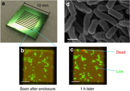
新たに開発されたX線自由電子レーザーは、フェムト秒という極めて短いパルス幅を持つため、主要な放射線損傷プロセスが起こる前の、ほとんど損傷のない試料を、シングルショット・スナップショットイメージングすることができる。生体イメージングでは、試料を自然に近い状態に保つことが不可欠である。しかし、従来の高分解能イメージングには、深刻な放射線損傷のため生きた細胞の可視化が妨げられるという欠点があった。今回我々は、微量液体封入アレイチップ内に保持された生きた細胞のスナップショットを、X線レーザー回折によって取得する方法を報告する。我々は、生きたMicrobacterium lacticum細胞を封入アレイチップに入れ、個々の封入領域に次々とSPring-8 Angstrom Compact Free-Electron Laserの単一X線レーザーパルスを照射した。封入チップ自体が保護スリットとなり、弱散乱体であるサブマイクロメートルサイズの細胞からの、28 nmの全周期分解能まで広がる明瞭なコヒーレント回折パターンが記録された。再構成された画像から、染色しない生きた細胞全体の構造が明らかになった。この手法は、細胞内現象の理解の深化に貢献する。
木村 隆志1, 城地 保昌2, 澁谷 明美3, Changyong Song3, Sangsoo Kim3, 登野 健介2, 矢橋 牧名3, 玉腰 雅忠4, 森屋 利幸5, 大島 泰郎5, 石川 哲也3, 別所 義隆3 & 西野 吉則1
- 北海道大学 電子科学研究所
- 高輝度光科学研究センター
- 理化学研究所 放射光科学総合研究センター
- 東京薬科大学 生命科学部
- 共和化工株式会社 環境微生物学研究所
Emerging X-ray free-electron lasers with femtosecond pulse duration enable single-shot snapshot imaging almost free from sample damage by outrunning major radiation damage processes. In bioimaging, it is essential to keep the sample close to its natural state. Conventional high-resolution imaging, however, suffers from severe radiation damage that hinders live cell imaging. Here we present a method for capturing snapshots of live cells kept in a micro-liquid enclosure array by X-ray laser diffraction. We place living Microbacterium lacticum cells in an enclosure array and successively expose each enclosure to a single X-ray laser pulse from the SPring-8 Angstrom Compact Free-Electron Laser. The enclosure itself works as a guard slit and allows us to record a coherent diffraction pattern from a weakly-scattering submicrometre-sized cell with a clear fringe extending up to a 28-nm full-period resolution. The reconstructed image reveals living whole-cell structures without any staining, which helps advance understanding of intracellular phenomena.