The Missing Kingdom: Why Fungi Must Be Central to Conservation Strategy
28 December 2025
Published online 16 December 2022
An analysis of a fly visual system reveals a small number of proteins that fundamentally define each distinct subtype of neuron.
Neset Ozel
Most key decisions during embryonic development are governed by transcription factor proteins, which bind to DNA and determine which genes get switched on or off. The resulting gene expression profile guides the fate of a stem cell as it matures into a fully functional adult cell. Researchers working with worm research models have identified ‘terminal selector’ transcription factors that act in different combinations to define neuronal identity early on, but no such factors have been firmly defined for other species to date.
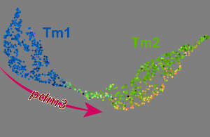
Desplan, along with postdoc Mehmet Özel, and NYU colleague Richard Bonneau , set out to uncover such terminal selectors in the fly visual system, which has a well-defined neuronal composition. “We know exactly which neurons are where, and what they do,” he says. His group has conducted systematic analyses of the gene expression activity in these cells, characterizing 200 specific neuronal types that collectively form the wiring for fly vision.
In their latest work, they computationally combed through these data to find patterns of transcription factor expression that consistently distinguish one neuronal type from another. Their analysis revealed that all this diversity arises through the differential expression of just 10 transcription factors, which are continuously expressed throughout cell maturation and drive the various subroutines associated with his development. And critically, Desplan’s team could experimentally validate these terminal selectors. “By changing this transcription factor that differs from one cell type to another, we should be able to reconfigure the cell type,” he says. “And it worked very well in this regard.”
Makoto Sato, a neurobiologist at Kanazawa University, in Japan who was not involved in the study, notes that the developmental process underlying cell type definition in the brain has long remained a black box. Desplan and Bonneau, he says, “have combined multiple cutting-edge technologies to expose what’s inside the black box using the fly brain as a model system.” Extending these findings to the mammalian brain will be far more challenging, but Desplan hopes that his colleagues in vertebrate biology will recognize the exciting future opportunities, including the potential to “make neurons almost from scratch”.
doi:10.1038/nmiddleeast.2022.80
Özel, M. et al. Coordinated control of neuronal differentiation and wiring by sustained transcription factors. Science https://dx.doi.org/10.1126/science.add1884 (2022).
28 December 2025
24 December 2025
24 December 2025
06 November 2020
15 January 2017
09 March 2016
Sign-up to receive our e-alert update every two weeks to keep up with everything new on the portal.
Sign up for e-alerts
Stay connected: